Cancer diagnosis without an invasive biopsy? Yes, please
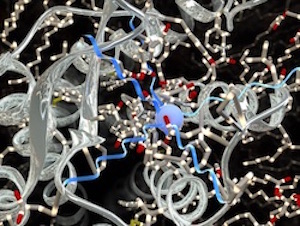
Radiolabelling is a technique that uses minute traces of radioactivity to track and locate tumor cells. Credit: Ella Maru Studio.
Is it cancer or is it a benign tumor? That question is often answered by invasive techniques like biopsies that involve needles or tubes to reach and remove a piece of tissue for closer study.
However, accurately diagnosing and treating cancer may soon be possible without a biopsy, thanks to researchers at Western University in London, Ontario.
A team led by Dr. Len Luyt, a professor with the departments of Chemistry and Oncology, has developed a new class of imaging agents targeting the prostate. Molecular imaging using trace amounts of radioactive metals or dye administered by injection or patch would provide a detailed picture of what is happening in the prostate, allowing for a more accurate detection of tumors.
“We are a synthetic chemistry laboratory, and our research program involves the design, preparation and evaluation of new compounds for the imaging and treatment of cancer,” says Luyt, who is also a Senior Scientist with the London Regional Cancer Program.
“Prostate cancer is one area where there is a need to more confidently diagnose a tumor and estimate the extent of the disease, to avoid over-treatment,” he continues. “Our goal is to create imaging agents that will improve treatment decisions by accurately detecting and localizing cancer, so we know if treatment is needed and where to direct it. This can have a real effect on the lives of prostate cancer patients.”
By adding radioactive metals into peptides that target tumours, cameras are able to locate a tumour without a biopsy.
Having his research group embedded within a cancer research lab helps bring to light what he calls “unmet clinical needs,” such as the need for a diagnosis without a biopsy. That close connection also came into play with a project related to breast and other cancers likely to metastasize, which previously had no known molecules that would target them.
Peptide receptors are located on the surface of tumor cells. Luyt’s colleague at the London Regional Cancer Program and Western Oncology, Biochemistry and Surgery professor, Dr. Eva Turley, discovered a new one called RHAMM (receptor for hyaluronan-mediated motility). This is a protein in humans that regulates cell movement and stem cell differentiation and is overexpressed in breast cancer.
Luyt and Turley received the 2016 WORLDiscoveries Vanguard Award for Innovator(s) of the Year for their work with RHAMM to selectively stimulate fat growth under the skin, moderate inflammation and reduce scarring by controlling the body’s own natural regenerative processes. Researchers continue to advance RHAMM-based technologies to find treatments for a variety of inflammatory conditions, including emphysema, arthritis and bronchopulmonary dysplasia, a form of chronic lung disease in premature infants.
Two approaches combine for effective results
Luyt’s team takes two approaches to creating new compounds for the imaging and treatment of cancer:
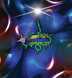
- On the chemistry side, the team works on technological advances for discovering compounds that target cancer. This includes methods to add radioactive metals into peptides that target tumors, which allows an external camera to view an image of the tumor.
- Applied projects with a specific cancer application, such as diagnosis or treatment related to prostate or breast cancer.
“The two areas of research really weave together,” Luyt says. “Ideally, the chemistry concepts we discover move into the application, resulting in improved methods of imaging and treatment.”
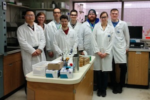
Professor Len Luyt, third from left, and the Western Faculty of Science graduate students are working to translate scientific discovery to clinical use.
Luyt’s team screens millions of different compounds to see which will interact with the cancer target, developing large “libraries” of peptides. Peptides play an important role in the body as hormones and neurotransmitters (relaying signals from one nerve cell to another).
Researchers don’t go directly to the library “shelf” for a specific peptide. “The starting point is typically a completely random situation,” Luyt explains. “As we discover compounds that target cancer, we create a focused library set up with portions of molecules we know are important, and start to make other changes to make the molecule even better.”
Luyt and Turley started a research project to find the peptides that would target RHAMM, discovering new molecules that bond well. The molecules show great potential as a drug and the team is working with a pharmaceutical company to move to clinical trials.
“This is an example of how chemists are able to collaborate with cancer scientists to move an academic biology discovery forward,” Luyt says.
Luyt appreciates the involvement of both undergraduate and graduate students from Western’s Faculty of Science, including its graduate program in molecular imaging, the only program of its type in Canada.
“People with an interest in imaging agents can directly interact with molecular biologists, physicists, other students and faculty,” he says. “They have a chance to see other pieces of the puzzle that will advance molecular imaging for cancer.”
He adds, “Chemists are not historically part of health research in Canada, but they play a key role in translating scientific discovery to clinical use. Specific applied projects may be more visible, but we need the fundamental science discoveries chemists can make to drive innovation, create new diagnostics and therapeutics for cancer and result in better outcomes.”
That includes fewer biopsies.