Consciousness and phantoms: how our brains compensate for an unexpected data gap
Imagine your brain as an airport.
In the airport, predictable networks get a passenger’s luggage checked in, get them through security, get them on the plane and coordinate the plane’s takeoff from a clear runway. Similarly, your brain’s networks communicate with each other so that you can simultaneously walk, talk and think about lunch.
But what happens when expected information isn’t there?
If everyone at the airport stops using one of the runways but no one tells the control tower, the tower will keep checking for activity without getting the responses it expects. The same thing happens in your brain, but while the airport situation would soon be fixed, it appears that the brain may sometimes react to missing information by “making it up”, in the form of phantom sensations such as pain in an amputated limb, or tinnitus in people with hearing loss.
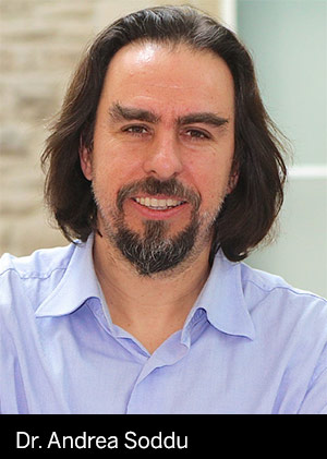
 New research may lead us closer to managing phantom sensation by helping us understand the origins. In a paper to be published this year by the prestigious medical journal The Lancet Neurology, Western University’s Andrea Soddu and an international team of researchers show that consciousness is a function of the way brain networks take turns sending and receiving information. If something happens to the brain that allows all information to be shared simultaneously – such as with epilepsy – consciousness is lost.
The connection to phantom sensation is that the brain anticipates this alternating pattern, and works to compensate when the pattern doesn’t happen as expected.
Soddu has a research focus on medical physics. He is a principal investigator at the Brain and Mind Institute and an assistant professor of physics and astronomy at Western. His lines of inquiry into consciousness and brain network function use St. Joseph’s Hospital’s combined PET-fMRI scanner.
How it works
PET (Positron Emission Topography) scans show glucose (energy) consumption by neurons, while fMRI (functional Magnetic Resonance Imaging) scans show blood flow to different parts of the brain in real time. By taking simultaneous scans, the researchers were able to see the networks in action, as well as the rate and patterns of energy consumption.
The focus of the study was a comparison of people in a vegetative state to people recovering from a “Minimally Conscious State”, also known as “MCS”. An MCS person is not vegetative, but is not able to control most of their thoughts and actions. They may be able to take a small deliberate action such as lifting a finger when asked to, but they can’t analyze questions and respond by tapping out a pattern. However, someone who is recovering or “exiting” MCS (“ExMCS”) may be able to reply in a pattern and eventually become able to communicate more effectively.
Soddu and his colleagues found that people in a vegetative state have “uncontrolled communication” between the two brain networks managing external-world input and internal thoughts – like a room full of people all shouting at once. The scans also confirmed that ExMCS patients retain some ability to use existing brain networks or compensate for them.
“When you look at vegetative or MCS people,” says Soddu, “their networks don’t take turns anymore; they are active together or silent together. However, in ExMCS, some awareness is there.”
Consciousness, networks and prediction
Soddu’s paper shares evidence that our brains actively seek information, like the control tower communicating with the runway. And this happens even when we’re not aware of thinking or doing something. In a relaxed and conscious but resting state – daydreaming, perhaps – our resting brains are active, seeking information and alerts that could affect us.
This constant seeking is like security guards looking for potential threats. Before the guards finds a threat, they will have considered other threat experiences and will have planned ahead so that they are ready to make the environment safe again as quickly as possible.
Our brains work the same way. They seek out and collect information and pair it with it memory – including millions of years of evolutionary development – and then plan ahead to keep us alive. In essence, our brains are built for prediction; for example, if we get hungry, we go to the kitchen because experience tells us that it is the most likely location for food.
Tinnitus
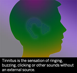
 Tinnitus is the sensation of ringing, buzzing, clicking or other sounds without an external source. Historically, it was believed that this was caused by damage to the delicate hairs of the inner ear, causing them to transmit incorrect information to the brain. However, other research by Soddu and his colleagues shows that the “sounds” are not created by the inner ear, but are likely created by the brain itself as a sort of prediction error. This could also apply to phantom limb pain experienced by amputees.
The research team made this connection by looking at the relaxed, conscious resting-state connectivity patterns in tinnitus patients. They observed that non-auditory regions of the brain were more active in patients experiencing episodes of tinnitus, while the auditory regions weren’t.
To put it simply, they saw that the brain compensates for the lack of expected real auditory input, and “makes up” sounds to fill the gap.
More knowledge, more potential
The findings on network patterns are major steps forward. But they do not yet offer any solutions for tinnitus or disorders of consciousness, only clues. However, more research began earlier this year with partnerships between Soddu’s team and Naples, Italy’s SDN-Istituto di Ricerca Diagnostica e Nucleare and the nearby neuro-rehabilitation center of the Salvatore Maugeri Foundation in Telese. The SDN also has a PET-fMRI machine, and together with the Western/St. Joseph’s researchers, they will be able to build a larger patient base with a wider range of symptoms. And that means better data, with more opportunities for knowledge and potential.
The Lancet Neurology paper, Neural correlates of consciousness in patients who have emerged from a minimally conscious state: a cross-sectional multimodal imaging study was authored by an international team including lead researcher Carol Di Perri of Belgium’s University of Lieges Coma Science Group, and Western University’s Andrea Soddu.
Funding for this research was provided by Belgian National Funds for Scientific Research (FNRS), European Commission, Natural Sciences and Engineering Research Council of Canada, James McDonnell Foundation, European Space Agency, Mind Science Foundation, French Speaking Community Concerted

