

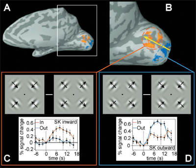
 The early visual areas of the brain such as area V1 are retinotopic. That is they show an orderly map of the visual world. Researchers have often used this map to interpret the relationship between fMRI activation and perception. They have assumed there is a one-to-one relationship between the layout of the retinotopic map and our perception of the world. Our research team (led by my former postdoc, David Whitney) have found that this is not necessarily the case. Using a motion-dependent visual illusion, which made stationary apertures containing a moving grating appear displaced from their real positions in the direction of motion, we showed that the activation in area V1 did not correspond to this change in apparent position. That is, although subjects' perceptions changed, there was no corresponding change in the map of activity in the brain. In essence, subjects perceived the object to be in one location but their brain represented the object as being in a completely different location. In fact, activation was greatest at the trailing edge of the moving gratings used in the display (and this was true even when the moving gratings were surrounded by a hard edge and did not show an illusory shift in position). These findings have important implications for conclusions drawn from the location of fMRI activation in retinotopic visual areas and also suggest that the connection between brain activity and perceptual awareness is more complicated than was originally thought.
Whitney, D., Goltz, H., Thomas, C.G., Gati, J., Menon, R., & Goodale, M.A. (2003). Flexible retinotopy: Motion dependent position coding in the visual cortex. Science, 382, 878-881.
Download pdf or Download pdf of supplemental material