
Brain Plasticity Following Hearing Loss and Restoration
About Dr. Steve Lomber
 Dr. Steve Lomber is a Professor in the Department of Physiology and Pharmacology in the Schulich School of Medicine and Dentistry and is jointly appointed in the Department of Psychology in the Faculty of Social Sciences. In addition, he holds an appointment as a principal investigator in the National Centre for Audiology in the Faculty of Health Sciences.
Hearing loss has profound effects on an individual’s quality of life. It is socially isolating, restricts opportunities in the work place, and facilitates cognitive decline in later life. In Canada, hearing loss is one of the most common birth defects, chronic disabilities, and complaints of the elderly. However, little is known about what occurs to the brain as a consequence of deafness. Dr. Lomber’s lab studies how the brain changes when hearing loss occurs in childhood or adulthood. Specifically, he studies auditory cortex in hearing individuals and changes that occur in auditory cortex following deafness.
The brain’s ability to change is known as “plasticity”. The plastic changes in the brain help deaf individuals enhance their ability to use their remaining senses, such as sight and touch. Understanding the natural limits of brain plasticity will assist in the development of methods designed to overcome these limitations and promote increased brain plasticity. This increase can lead to improved success in the use of hearing restoration devices such as cochlear implants.
Cochlear implants are extremely useful tools in providing sound sensations to deaf individuals as well as making speech comprehension possible. Cochlear implants have restored hearing to hundreds of thousands of people around the world. Revealing the plasticity of the cerebral cortex after cochlear implantation will make it possible to develop therapeutic strategies to better serve the needs of the brain, enhance the benefits of cochlear implants, and improve hearing restoration success in children and the aged.
Study Results
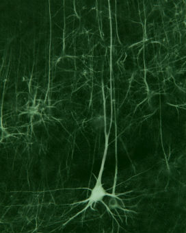 When the brain is deprived of input from one sensory modality, it often compensates with supranormal performance in one or more of the intact sensory systems. In the absence of acoustic input, it has been proposed that cross-modal reorganization of deaf auditory cortex may provide the neural substrate mediating compensatory visual function. By using a battery of visual psychophysical tasks we found that congenitally deaf, compared to hearing, cats have superior localization in the peripheral field and lower visual movement detection thresholds. Furthermore, in the deaf cats, reversible deactivation of posterior auditory cortex selectively eliminated superior visual localization abilities while deactivation of the dorsal auditory cortex eliminated superior visual motion detection. Taken together, this combination of experimental approaches demonstrates a causal link between the crossmodal reorganization of auditory cortex and enhanced visual abilities of the deaf, as well as identified the cortical regions responsible for adaptive supranormal vision.
Research Approach
 The long-term goal is to understand and define the capabilities of the auditory cortex to establish acoustic function following cochlear implant. By understanding how the cerebral cortex can adapt to process signals generated by a cochlear prosthetic, it will be possible to alter cochlear prosthetics to better serve the needs of the cerebrum. This endeavor requires four steps: 1) The elucidation of the essential contributions that primary and non-primary auditory cortex make to fundamental acoustically-guided behaviors in the hearing population, 2) A determination of the acoustic abilities of deaf subjects following cochlear implant, 3) An assessment of the behavioral capabilities of primary and non-primary auditory cortex following cochlear implant in deaf subjects, and 4) An examination of the plastic changes that can be made by the auditory cortex when the age at time of cochlear implant is altered. To that end, we examine auditory cortical function in the hearing animals, deaf animals, and animals following cochlear implant.
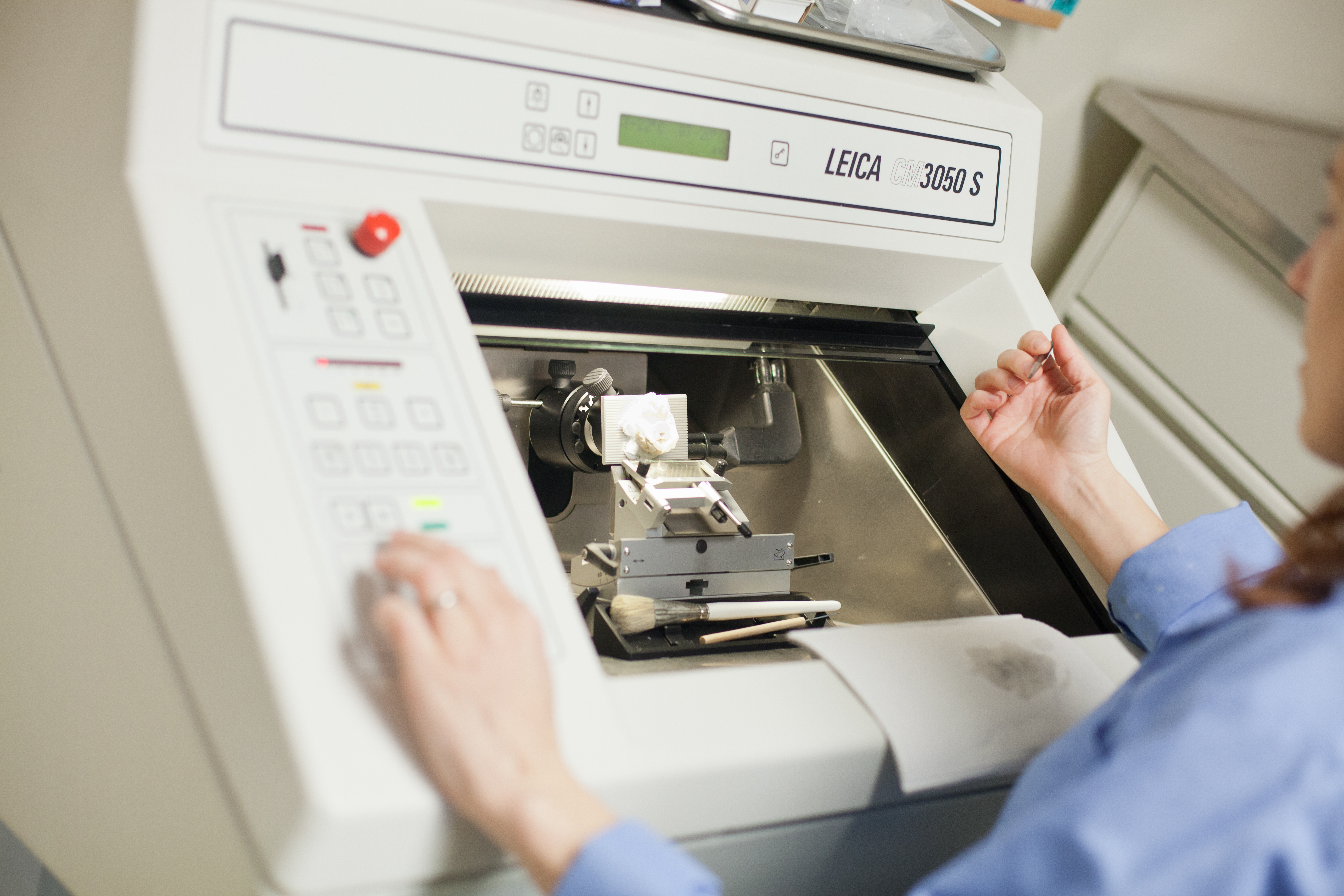
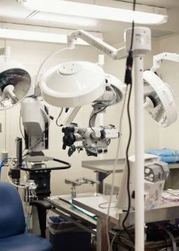
 The Cerebral Systems Lab uses an integrative approach to examine cortical plasticity following hearing loss and restoration. This approach includes psychophysical testing, electrophysiological recording, connectional anatomy, reversible deactivation, and functional imaging.
References
Butler, B.E. and Lomber, S.G. (2013) Functional and structural changes throughout the auditory system following congenital and early-onset deafness: implications for hearing restoration. Frontiers in Systems Neuroscience 7:92. pgs. 1-17.
Brown, T.A., Joanisse, M.F., Gati, J.S., Hughes, S.M., Nixon, P.L., Menon, R.S. and Lomber, S.G. (2013) Characterisation of the blood-oxygen level-dependent (BOLD) response in cat auditory cortex using high-field fMRI. NeuroImage 64: 458-465.
Meredith, M.A., Kryklywy, J., McMillan, A.J., Malhotra, S., Lum-Tai, R. and Lomber, S.G. (2011) Crossmodal reorganization in the early-deaf switches sensory, but not behavioral roles of auditory cortex. Proceedings of the National Academy of Sciences (USA) 108: 8856-8861.
Lomber, S.G., Meredith, M.A. and Kral, A. (2010) Crossmodal plasticity in specific auditory cortices underlies visual compensations in the deaf. Nature Neuroscience 13: 1421-1427.
Lomber, S.G. and Malhotra, S. (2008) Double dissociation of “what” and “where” processing in auditory cortex. Nature Neuroscience 11: 609-616.